The Current Research Projects
of Prof. Zhenyu Guo
at the
Laboratory of Biomedical Engineering
Project1: Three-dimensional Power Doppler Angiography
(Click on images for larger views)
 The therapeutic options for patients having lower-extremity arterial occlusive
diease requires vascular imaging that can accurately quantify the severety of
stenosis and reveal all distall runoff vessels suitable for bypass grafting.
Currently, contrast angiography is the standard technique for this purpose.
However, this invasive technique has failed to demonstrate suitable distall
vessels for reconstructive surgery in up to 70% of patients with severe
occlusive diease. Although duplex ultrasound scanning is today the most widely used noninvasive technique to quantify arterial stenoses, recent studies showed that there are significant limitations to use it in situations invloving
multiple stenoses. Magnetic resonance angiography has been considered as a
potential technique to replace contrast angiography, but its avaiability to
accurately diagnose the arterial occlusive disease is limited by the signal loss due to flow turbulence encountered in the stenotic jets.
The therapeutic options for patients having lower-extremity arterial occlusive
diease requires vascular imaging that can accurately quantify the severety of
stenosis and reveal all distall runoff vessels suitable for bypass grafting.
Currently, contrast angiography is the standard technique for this purpose.
However, this invasive technique has failed to demonstrate suitable distall
vessels for reconstructive surgery in up to 70% of patients with severe
occlusive diease. Although duplex ultrasound scanning is today the most widely used noninvasive technique to quantify arterial stenoses, recent studies showed that there are significant limitations to use it in situations invloving
multiple stenoses. Magnetic resonance angiography has been considered as a
potential technique to replace contrast angiography, but its avaiability to
accurately diagnose the arterial occlusive disease is limited by the signal loss due to flow turbulence encountered in the stenotic jets.
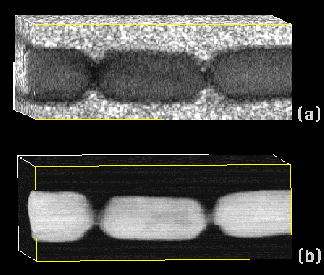
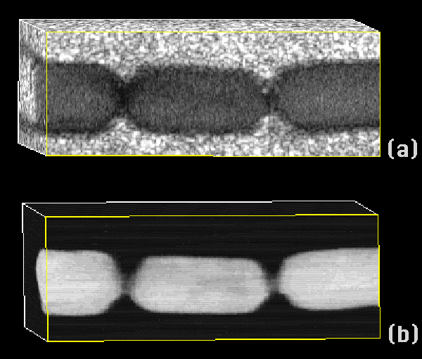
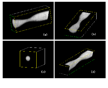
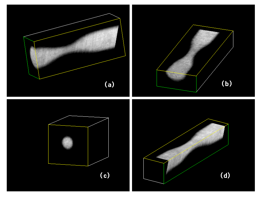
In this project, we use three-dimensional (3D) power Doppler angiography (PDA)
for quantifying lower-extremity arterial stenoses and revealing distall runoff
vessels. This technique has high sensitivity to map flowing blood. It is
nearly flow velocity and Doppler angle independent and essentially alsising-
free. With this technique, physicians should be able to analyze the lower-
extremity arteries for anatomic and pathological purpose in 3D. This will
significantly increase the accuracy for quantifying stenoses and facilitate the salvaging limbs surgery planning. we have done substantial research on 3D PDA. Attached figures show some of our 3D power Doppler angiograms.
The general objective of the current project is to extend 3D PDA to routine
clinical practice by improving the current mothod. This project will result
in a clinical relevant 3D PDA system for quantifying lower-extremity arterial
stenoses and revealing run-off vessels. Successfully completion of this project will produce a great impact on reducing the health care cost on cardiovascular disease as this technique can be expended to diagnose stenoses of other arterial beds, to monitor bypass graft patency, and to diagnose deep vein thrombosis.
Project2: Medical Virtual Instrumentation
In normal clinical practice, several medical instruments are required for the
measurement and monitoring of various physical and physiological data. These
instruments function as single, stand-alone devices capable of performing
individual tasks and lack the flexibility required to perform multiple tests and kinds of monitoring. In addition, small medical clinics may not have all the
required instruments, meaning that patients have to be referred to medical
centers for simple physiological tests. It is thus of great value to
develop a general purpose instrument with the cost of a single-functional
instrument which can measure different biomedical data.
The current clinically applied monitoring instruments mainly perform simple
monitoring and measurement functions, without the ability to perform advanced
signal processing to extract valuable diagnostic information. In addition,
conventional medical instruments do not perform remote(i.e.. tele-medicine)
measurements or be controlled remotely. It is thus of great value to develop a
medical instrument which has the abilities to conduct advanced signal
processing for biopotentials and to perform remote measurements.
The general objectives of this medical virtual instrumentation research are:
1- to embed the functions of various medical monitoring instruments in a
single location, the personal computer, by using virtual instrumentation
technology to develop a multi-functional medical virtual instrument; 2- to
integrate advanced signal processing techniques into this medical virtual
instrument to facilitate the extraction of diagnostic information; and 3- to
connect this medical virtual instrument to the World Wide Web (WWW) and
remotely control this instrument from any Web Browser for tele-medicine
practice and home medical care delivery.
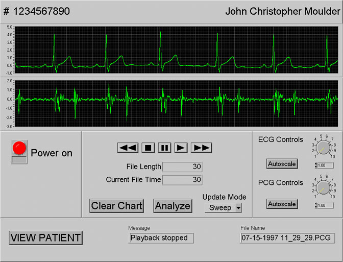
Through the use of suitable transducers, this instrument can be used to monitor and measure physiological data such as blood pressure, heart sounds, respiratory flow, body temperature, muscle force as well as many other parameters. It can also be used with electrodes for monitoring and diagnosing biopotentials such as electrocardiogram (ECG), the electromyogram (EMG), and the electroencephalogram (EEG). Such an instrument is also ideal for physiology education. This
medical virtual instrument include a micro-controller controlled multi-channel
signal conditioner, plug-in data acquisition boards, software modules used to
create instrument front panels on the computer monitor with which to conduct
various tasks, as well as algorithms used to perform signal processing and
Internet communication.
Comparing with the conventional medical instruments, our instrument has the
following advantages:
1- Patient management. Since a PC is the core of the instrument, patients can
be managed using patient data base.
2- Reduce the health care cost. Multiple functional virtual instrument will
have a cost of a stand-alone instrument due to the fact that software is the
instrument.
3- Capture and display current signals while saving or analyzing the previous
signals using inherent multiprocessing of the Pentium PC. This is very
important as the traditional instrument can only perform one task at a time.
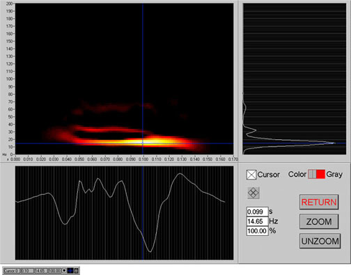
4- Advanced signal processing can be performed to the medical data recorded.
For example, modern spectral analysis techniques and time-frequency analysis
can be applied to extract useful diagnostic information which is not possible in the traditional instruments.
5- A key function of this instrument is the ability to connect to the Internet. All front panels of this instrument are viewable and controllable from any
browser via the Internet, providing a means to deliver home medical care.
In summary, this project will provide the most advanced and cost-effective
technology for medical instrumentation. The multi-functional medical
virtual instrument can be used in routine clinical pratice and tele-medicine,
it can also be used in physiological and biomedical engineering laboratories
for teaching and research.
Project3: Three-dimensional Image Compression using Iterated Function
Systems (under construction)

Return to Zhenyu's home page
 The therapeutic options for patients having lower-extremity arterial occlusive
diease requires vascular imaging that can accurately quantify the severety of
stenosis and reveal all distall runoff vessels suitable for bypass grafting.
Currently, contrast angiography is the standard technique for this purpose.
However, this invasive technique has failed to demonstrate suitable distall
vessels for reconstructive surgery in up to 70% of patients with severe
occlusive diease. Although duplex ultrasound scanning is today the most widely used noninvasive technique to quantify arterial stenoses, recent studies showed that there are significant limitations to use it in situations invloving
multiple stenoses. Magnetic resonance angiography has been considered as a
potential technique to replace contrast angiography, but its avaiability to
accurately diagnose the arterial occlusive disease is limited by the signal loss due to flow turbulence encountered in the stenotic jets.
The therapeutic options for patients having lower-extremity arterial occlusive
diease requires vascular imaging that can accurately quantify the severety of
stenosis and reveal all distall runoff vessels suitable for bypass grafting.
Currently, contrast angiography is the standard technique for this purpose.
However, this invasive technique has failed to demonstrate suitable distall
vessels for reconstructive surgery in up to 70% of patients with severe
occlusive diease. Although duplex ultrasound scanning is today the most widely used noninvasive technique to quantify arterial stenoses, recent studies showed that there are significant limitations to use it in situations invloving
multiple stenoses. Magnetic resonance angiography has been considered as a
potential technique to replace contrast angiography, but its avaiability to
accurately diagnose the arterial occlusive disease is limited by the signal loss due to flow turbulence encountered in the stenotic jets.